The Laboratorio de Optica in the University of Murcia (LOUM) is one of the pioneer groups worlwide in applying Adaptive Optics in the study of the eye and it has obtained an important number of fundamental results on the optics characteristics of the eye and the retina. The research group was founded in 1994 with the aim of becoming a reference in the world. Nowdays is a solid and consolidated group with 17 members and that recently moved to the new facilities in the Centro de Investigación en Óptica y Nanofísica (CiOyN) building in the University of Murcia.
Images on the right show a recent picture of the research group members and the new building.
Parallel to the developments in basic research, the group is in permanent relation with national and international companies in Opthalmic Optics. Indeed, some of the new instruments and opthalmic products currently in the market have been developed in our lab. Now, we describe very briefly some of the contributions with a higher impact in the fields of:
- Visual Optics
- Adaptive Optics
And also we mention some of the currents projects in our laboratory.
Visual Optics
The main goal of our laboratory has been to develop new instrumentation to better understand the optics of the human eye and the retina. Firstly, we centered our activity on the study of systems and instruments to evaluate the retinal image quality and to measure aberrations (an asymmetric double-pass which allowed to estimate the retinal image of a point -PSF-), and on the use of phase-retrieval algorithms to calculate the wavefront aberration [1-3]. The commercially available instrument OQAS (Visiometrics, S.L.) is based on these studies. Afterwards, we built one of the first Hartmann-Shack wavefront sensors [4]. This was later modified in collaboration with the University of Rochester in order to measure aberrations in real time at 25 Hz [5].
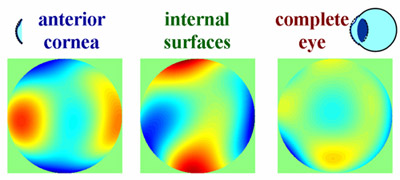
Example of compensation of the aberrations between the cornea and the lens
On the study of sources and location of ocular aberrations, LO·UM has reported some new results along the last decade. By comparing corneal aberrations (computed from elevation data [6]) and the whole eye, we separated the contribution of the different ocular components. We reported that the lens might compensate for most of the first-surface corneal aberrations [7,8]. More recently, we have obtained some understanding on the mechanism responsible for this compensation [9], and we believe this is an automatic procedure more significant in eyes presenting larger angles, that is hiperopic eyes. Ocular optics also change with age. One of our lines of research tried to know the reasons for these age-related changes. We found that the quality of the retinal image decreases with age [10] but the corneal optics remains almost constant [11].This means that this reduction is due to a decrease in the optical quality of the lens [12], and more specifically to a decoupling of the corneal and lens aberrations.
The paper reporting this results is highly cited (with nearly 100 citations is the top-1 paper cited in JOSAA for the last 5 years). These basic science results have been the origin for the development of a new generation of intraocular lenses with negative spherical aberration [13]. Now this type of lenses (TECNIS lens by AMO was the first IOL of this type) is world-wide accepted. The polarization properties of eye and the retina and their effect on the retinal image quality estimates have also been study in depth. Some polarimetric experimental setups were developed which might be the origin of future clinical instrument [14-17]. Finally, we have also been involved in experiments closely related to Ophthalmic Optics. As an example, the work developed in collaboration with ESSILOR International and centered on the analysis of the optical properties of progressive power lenses [18,19] has been used for the development of a new generation (and recently out in the market) of progressive power lenses: Varilux Physio.
Adaptative Optics
Once robust systems to measure aberrations were developed, the progress towards the correction was natural. Adaptive Optics (AO) techniques, first developed for astronomical purposes, allowed to increase the resolution of images in telescopes (limited by the atmospheric turbulence) by compensating the wavefront. The high cost of this technology limited its use to astronomical purposes and military applications. Our laboratory was one of the first to propose a static correction of the ocular wavefront aberrations. We initially used a liquid-crystal spatial modulator and reported results in the human eye limited by the characteristics of the corrector (number of elements and stroke), but we showed potential applications with deformable mirrors [20].
Lately we decided to use a membrane deformable mirror. The price was one order of magnitude below that of ‘classical’ deformable mirrors and we thought to be suitable for applications in the human eye. We reported the first correction for ocular aberrations in close-loop [21].
This prototype was used to improve the AO experiments carried out in our laboratory. In particular, a second generation of ocular AO using two correction devices simultaneously (a membrane deformable mirror [22] and a spatial liquid-crystal crystal modulator with XGA resolution [23]) were also reported. This system, called “AO visual simulator”, has been mainly used to study the relationship among the ocular optics, accommodation and visual quality [24-26].We have shown the large flexibility of AO setups in Visual Optics applications. Image below shows the existing system in 2005.
Image of the first adaptive optics prototype working in close loop and real time (for more details, see ref 21).
AO visual simulator with the spatial modulator in 2005.
Another application of AO is the improvement of retinal images. More than 10 years ago, Artal and Navarro [27] proposed the use of high resolution techniques (previously used in Astronomy) in the human eye. In 1997, we obtained retinal images with a high magnification system, although just with partially-corrected aberrations [20]. We also applied our technique to extended images which gave good results [28] and was lately used by others. In parallel, we also explored an alternative way of imaging the retina based on a scanning laser system [29].
The incorporation of AO techniques into conventional retinal imaging setups increases lateral resolution in the best in-focus plane. However, axial resolution is low. One solution is to use low coherence tomographic techniques (OCT). In this topic we have been collaborated with W. Drexler’s group (formerly at the University of Vienna) in the last years. This collaboration allowed us to get the first OA topography retinal images [30].
References:
[2] I.Iglesias, N.Lopez and P. Artal, “Reconstruction of the ocular PSF from a pair of double pass images by phase retrieval techniques”, J.Opt.Soc.Am.A. 15, 326-339 (1998).
[3] I.Iglesias, E.Berrio and P.Artal, “Estimates of the ocular wave aberration from pairs of double pass retinal images”. J.Opt.Soc.Am.A. 15, 2466-2476 (1998).
[4] P.M. Prieto, F. Vargas, S. Goelz and P.Artal, ” Analysis of the performance of the Hartmann-Shack sensor in the human eye” J.Opt.Soc.Am.A. 1:1388-98 (2000).
[5] H. Hofer, P.Artal, B.Singer, J.L.Aragon, D. R. Williams, “Dinamics of the eye wave aberration”, J.Opt.Soc.Am.A.18, 497-506 (2001).
[6] A.Guirao and P.Artal. “Córneal wave-aberrations from videokeratography: accuracy and limiations of the procedure” J.Opt.Soc.Am.A. 17, 955-965 (2000).
[7] P.Artal and A.Guirao “Contribution of córneal and lns to the aberrations of the human eye”. Opt.Lett. 23, 1713-1715 (1998).
[8] P. Artal, A. Guirao, E. Berrio, & D.R. Williams, “Compensation of corneal aberrations by the internal optics in the human eye”. Journal of Vision, 1(1), 1-8, (2001).
[9] P. Artal, A. Benito, J. Tabernero , “The human eye is an example of robust optical design”, J. Vis. , 6 , 1–7 (2006).
[10] A.Guirao, C.Gonzalez, M.Redondo, E.Geraghty, S.Norrby and P.Artal. “Average optical performance of the human eye as afunction of age in a normal population”, Inv.Oph.Vis.Sci. 40, 203-213 (1999).
[11] A.Guirao and P.Artal. “Córneal wave-aberrations from videokeratography: accuracy and limiations of the procedure”. J.Opt.Soc.Am.A. 17, 955-965 (2000).
[12] P. Artal, E. Berrio , A. Guirao and P. Piers. “Contribution of the cornea and internal surfaces to the change of ocular aberrations with age” J. Opt. Soc. Am. A, 19 , 137-143 (2002).
[13] A. Guirao, M. Redondo, E. Geraghty, P. Piers, S. Norrby and P. Artal. “ Corneal optical aberrations and retinal image quality in patients in whom monofocal intraocular lenses were implanted” Arch Ophthalmol., 120,1143-51 (2002).
[14] J.Bueno and P.Artal. “Double-pass imaging polarimeter in the eye” Opt.Lett. 24, 64-66 (1999).
[15] J.Bueno and P.Artal “Polarization and retinal image quality estimates in the human eye”. J.Opt.Soc.Am.A. 18, 489- 496 (2001).
[16] J. Bueno, E. Berrio and P. Artal, “Aberro-polariscope for the human eye” Opt.Lett. 2, 1209-1211 (2003).
[17] J .Bueno, E.Berrio, P.Artal, “Corneal polarimetry after LASIK refractive surgery”, J. Bio. Opt , 11 , (2006).
[18] E. A. Villegas, C. Gonzalez, B. Bourdoncle, T. Bonnin and P. Artal. “ Correlation between optical and psychophysical parameters as a function of defocus.” Optom. Vis Sci. 79(1):60-7. (2002).
[19] E. A. Villegas and P. Artal, “Spatially resolved wavefront aberrations of ophthalmic progessive-power lenses in normal viewing conditions” Optom. Vis. Sci., 80 , 106-114 (2003).
[20] F.Vargas, P.Prieto and P.Artal “Correction of the aberrations in the human eye with a liquid crystal spatial light modulator: limits to the performance” J.Opt.Soc.Am.A. 15, 2552-2562 (1998).
[21] E.Fernández, I.Iglesias, P.Artal “Closed loop adaptive optics in the human eye”.Opt.Lett.26, 746-748 (2001).
[22] E. Fernández and P. Artal, “Membrane deformable mirror for adaptive optics: performance limits in visual optics” Opt. Express, 11 , 1056-1069 (2003).
[23] P. M. Prieto, E. J. Fernández, S. Manzanera and P. Artal, “Adaptive optics with a programmable phase modulator: applications in the human eye” Opt. Express , 12 , 4059-4071 (2004).
[24] P. Artal, L. Chen, E. J. Fernández, B. Singer, S. Manzanera and D. R. Williams, “Neural adaptation for the eye’s optical aberrations” J. Vis. , 4 , 281-287 (2004).
[25] E. J. Fernández and P. Artal, “Study on the effects of monochromatic aberrations in the accommodation response by using adaptive optics”, J. Opt. Soc. Am. A , 22 , 1732-1738 (2005).
[26] P. A. Piers, E. J. Fernández, S. Manzanera, S. Norrby and P. Artal, “Adaptive optics simulation of intraocular lenses with modified spherical aberration” Invest. Ophthalmol. Vis. Sci. , 45 , 4601-4610 (2004).
[27] P.Artal and R.Navarro. “High-resolution imaging of the living human fovea: measurement of the intercenter cone distance by speckle interferometry” Optics Letters, 14, 1098-1100 (1989).
[28] I.Iglesias & P.Artal “Deconvolution of retinal images from wave -front data”. Opt.Lett. 25,1804-1806 (2000).
[29] B. Vohnsen, I. Iglesias & P. Artal, “Directional Imaging of the Retinal Cone Mosaic” Opt. Lett. , 29 , 968-970 (2004).
[30] B. Hermann, E. J. Fernández, A. Unterhuber, H. Sattmann, A. F. Fercher, W. Drexler, P. M. Prieto and P. Artal, Opt. Lett. , 29 , 2142-2144 (2004).