Abstract
Purpose:
To provide a comprehensive investigation of the optical quality across the visual field for current mainstream types of refractive surgeries.
Methods:
Sixty eyes from 60 adults who received refractive surgery of either femtosecond laser–assisted laser in situ keratomileusis (FS-LASIK), Q-value guided customized laser in situ keratomileusis (Q-LASIK), small incision lenticule extraction (SMILE), or Implantable Collamer Lens (ICL) (STAAR Surgical) implantation were included in this study. Refraction and optical aberrations from a visual field of horizontal 60° (from temporal 30° to nasal 30°) and vertical 36° (from superior 20° to inferior 16°) were measured using a custom-made Hartmann-Shack wavefront peripheral sensor. Refractive error, higher order aberrations, point spread function (PSF), and Strehl ratio were compared among these groups prior to and after the surgical procedures, respectively.
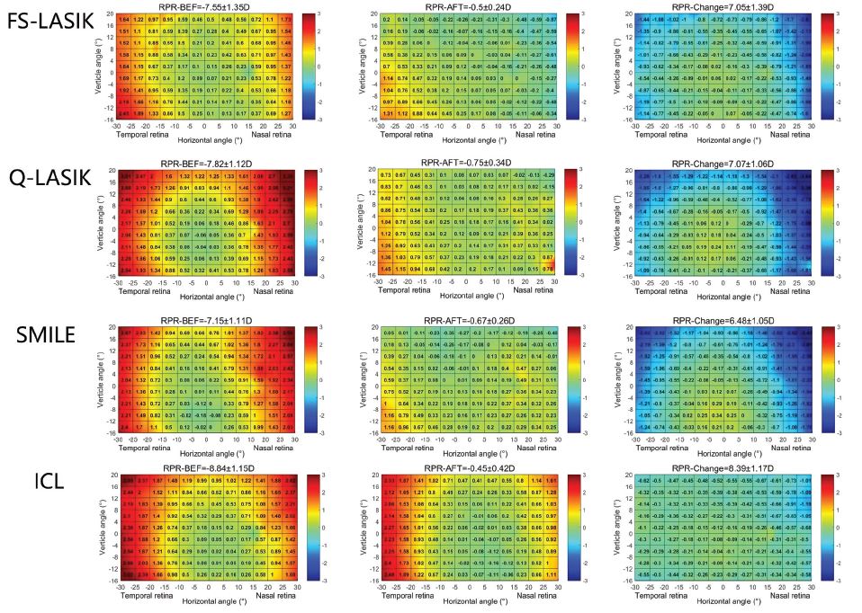
Results:
All types of surgical procedures achieved an almost plano refraction in the central retina. This was also the case in the peripheral retina for the three types of laser refractive surgeries. However, residual peripheral relative hyperopic defocus was observed after ICL implantation. In all groups prior to the surgery, PSFs showed increasing distortion with eccentricity and arrow-like shape pointing toward the central fovea in the periphery in diagonals. Degradation of the PSFs was diminished by all three types of laser refractive surgeries, whereas ICL implantation made the peripheral distortion more prominent.
Conclusions:
Although ICL implantation produced a similar impact on refractive correction and objective optical quality in the central vision compared with other laser refractive surgeries, its outcome on the peripheral optics is different. The impact of this difference on visual performance deserves notice and warrants further investigation.
 0
0 0
0