Visualizing In Vivo Human Ocular Tissues with Two-Photon Microscopy
Overview
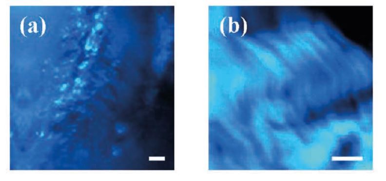
Visualization and analysis of transparent living ocular tissues at microscopic scale have been a challenge during the last decades. Nowadays clinical devices only allow imaging cellular corneal structures; however, collagen fibers of the stroma (90 % of the corneal thickness) cannot be imaged. It has been recently reported a compact Second Harmonic Generation (SHG) microscope able to image the living human cornea and the sclera for the very first time.
If you like it, please share it... 0
0 0
0
00