Purpose: To analyze the spatial organization of pathological corneas with second harmonic generation (SHG) imaging and to provide a proof of concept to objectively distinguish these from the healthy corneas.
Methods: A custom-built SHG microscope was used to image the anterior stroma of ex vivo corneas, both control and affected by some representative pathologies. The structure tensor (ST) was employed as a metric to explore and quantify the alterations in the spatial distribution of the collagen lamellae.
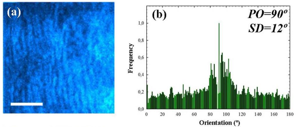
Results: The collagen arrangement differed between healthy and pathological samples. The former showed a regular distribution and a low structural dispersion (SD , 408) within the stroma with a well-defined dominant orientation. This regular arrangement drastically turns into a disorganized pattern in pathological corneas (SD. 408).
Conclusions: The combination of SHG imaging and the ST allows obtaining quantitative information to differentiate the stromal collagen organization in healthy and diseased corneas. This approach represents a feasible and powerful technique with potential applications in clinical corneal diagnoses.
Translational Relevance: The ST applied to SHG microscopy images of the corneal stroma provides an experimental objective score to differentiate control from pathological or damaged corneas. Future implementations of this technique in clinical environments might might be a promising tool in Ophthalmology, not only to diagnose and monitor corneal diseases, but also to follow-up surgical outcome.
DOI: https://doi.org/10.1167/tvst.8.3.51
 0
0 0
0