Abstract
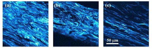
Papillary carcinoma is the most prevalent type of thyroid cancer. Its diagnosis requires accurate and subjective analyses from expert pathologists. Here we propose a method based on the Hough transform (HT) to detect and objectively quantify local structural differences in collagen thyroid nodule capsules. Second harmonic generation (SHG) microscopy images were acquired on non-stained histological sections of capsule fragments surrounding the healthy thyroid gland and benign and tumoral/malignant nodules. The HT was applied to each SHG image to extract numerical information on the organization of the collagen architecture in the tissues under analysis. Results show that control thyroid capsule samples present a non-organized structure composed of wavy collagen distribution with local orientations. On the opposite, in capsules surrounding malignant nodules, a remodeling of the collagen network takes place and local undulations disappear, resulting in an aligned pattern with a global preferential orientation. The HT procedure was able to quantitatively differentiate thyroid capsules from capsules surrounding papillary thyroid carcinoma (PTC) nodules. Moreover, the algorithm also reveals that the collagen arrangement of the capsules surrounding benign nodules significantly differs from both the thyroid control and PTC nodule capsules. Combining SHG imaging with the HT results thus in an automatic and objective tool to discriminate between the pathological modifications that affect the capsules of thyroid nodules across the progressions of PTC, with potential to be used in clinical settings to complement current state-of-the-art diagnostic methods.
© 2020 Optical Society of America
 0
0 0
0